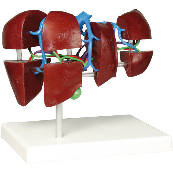
This reduced size model shows the division of the liver into 8 liver segments after C. Couinaud. It explains the separate vessel supply of the individual segments. The distribution of the portal vein separates the liver in a transverse plane into an upper (cranial) and lower (caudal) Segment group.
The following segments are depicted:
Left Liver Lobe
- Segment I caudate lobe
- Segment II cranial part of lateral segment
- Segment III caudal part of lateral segment
- Segment IV quadrate lobe
- Segment IVa cranial part
- Segment IVb caudal part
- Right Liver Lobe
- Segment V caudal part of anterior segment
- Segment VI caudal part of posterior segment
- Segment VII cranial part of posterior segment
- Segment VIII cranial part of anterior segment
The model is not dissectible and comes on a removable stand.
Size: 18 x 13 x 17 cm, weight: 0.5 kg

